Biomedical Imaging
The Biomedical Imaging Core (BIC) is a state-of-the-art research facility dedicated to advancing the study of cognition and biological processes through non-invasive imaging techniques. Our core provides cutting-edge methodologies for exploring human brain function in neuroscience and psychology research while also supporting comprehensive studies of anatomical, functional, and physiological processes in animal models.
To support human cognitive research, we employ multiple imaging modalities, including functional magnetic resonance imaging (fMRI), magnetoencephalography (MEG), and electroencephalography (EEG). By synchronizing these technologies with visual and/or auditory stimuli and coordinating their recordings with motor responses, we can precisely map the relationship between neuronal activity and behavior across both time and space. This multimodal approach enhances the resolution of cognitive function studies, neural dynamics, and sensory processing, offering a more comprehensive understanding of brain function than any single modality alone. It enables researchers to investigate the complex interplay of neural activity across different scales, providing deeper insights into brain mechanisms.
For preclinical research, our facility offers advanced small animal imaging technologies to facilitate non-invasive studies on disease progression, drug efficacy, and molecular biological processes in rodents. These imaging tools provide high-resolution insights into structural and physiological dynamics, improving the accuracy and depth of preclinical research. The facility is equipped with bioluminescence and fluorescence imaging for capturing cellular and mesoscopic resolution processes, a micro-CT scanner for detailed anatomical assessments, and endoscopic imaging for direct tissue visualization, biopsy, and targeted injections.
A key strength of the Biomedical Imaging Core is its ability to integrate multiple imaging modalities within a single study, enhancing the accuracy and depth of research findings. By leveraging cutting-edge imaging technologies across multiple research domains, we enhance scientific collaboration and accelerate discoveries in human cognition, disease mechanisms, and therapeutic interventions. This interdisciplinary model fosters novel methodologies and drives progress in both fundamental and applied biomedical research.
Equipment List
1. Brain Imaging Lab (BI)
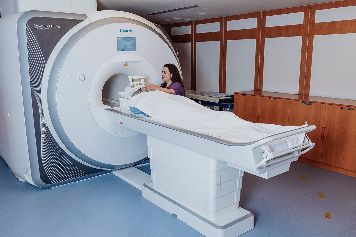
The Brain Imaging Lab focuses on the non-invasive examination of structural and functional aspects of the human body, with applications in neurology, cognitive science, and clinical research. Using advanced MRI technology combined with simultaneous EEG recording, researchers can study physiological processes with high spatial and temporal resolutions.
Main Equipment
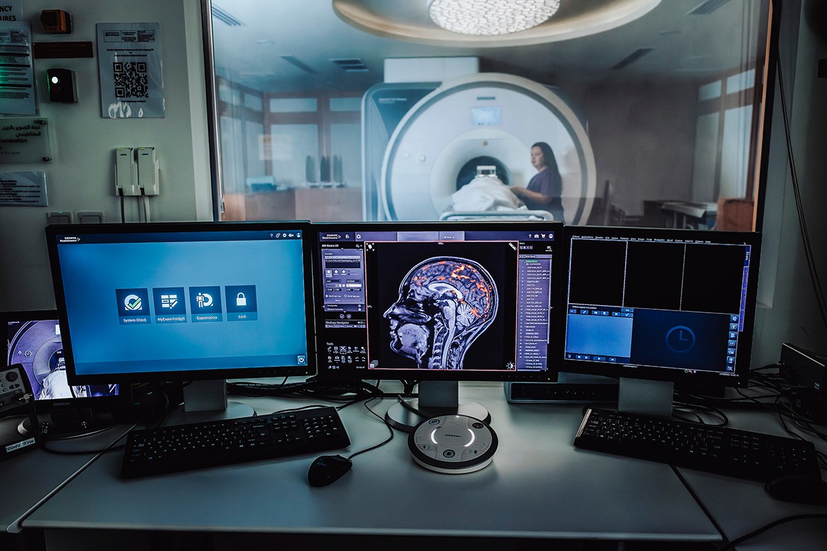
Siemens MAGNETOM Prisma 3T MRI scanner with advanced pulse sequences including simultaneous multi-slice (SMS) for BOLD and diffusion imaging for enabling high spatial and temporal resolution images
- Multiple RF coils (20, 32, and 64-channel head "TIM," and body coils)
Supporting Equipment
- Eye Link 1000 Eye Tracking System
- Vpixx Projector System for visual and auditory stimuli and response recording
- MR-compatible digital video camera (MRC-HiSpeed) and microphone
- ResponsePixx 5-button fiber-optic Response box for motor response synchronization
- 64 Channel Brain Products EEG-fMRI compatible System for simultaneous EEG and fMRI acquisition

2. NeuroWaves Lab (NW)
The NeuroWaves Lab specializes in measuring neural activity using magnetoencephalography (MEG) to detect magnetic fields generated by neural currents and electroencephalography (EEG) to record electrical brain activity. This lab provides insights into neural dynamics, sensory processing, and cognitive research applications.
Main Equipment
- Kanazawa Institute of Technology (KIT) MEG System with 208 axial gradiometers for high spatio-temporal resolution recording of local neural activity
- OPM MEG by Fieldline with 96 channel system
- EEG System by Brain Products with 32/64 channel for high spatio-temporal resolution recording of brain electrical activity
Supporting Equipment
- FastSCAN Laser Scanner for precise anatomical mapping and source localization
- Eye Link 1000 Eye Tracking System for synchronized gaze tracking
- Vpixx Projector System for sensory stimulation and response recording
- ResponsePixx 5-button fiber-optic Response box for motor response synchronization

3. Small Animal Imaging Lab (SAI)
The Small Animal Imaging Lab, located within the AAALAC-accredited vivarium, offers multimodal imaging capabilities for preclinical research, supporting investigations into disease progression, therapeutic interventions, and molecular biology in animal models.
Main Equipment
- Perkin Elmer (Revvity) IVIS Spectrum Imaging System for in vivo 2D and 3D bioluminescence and fluorescence imaging.
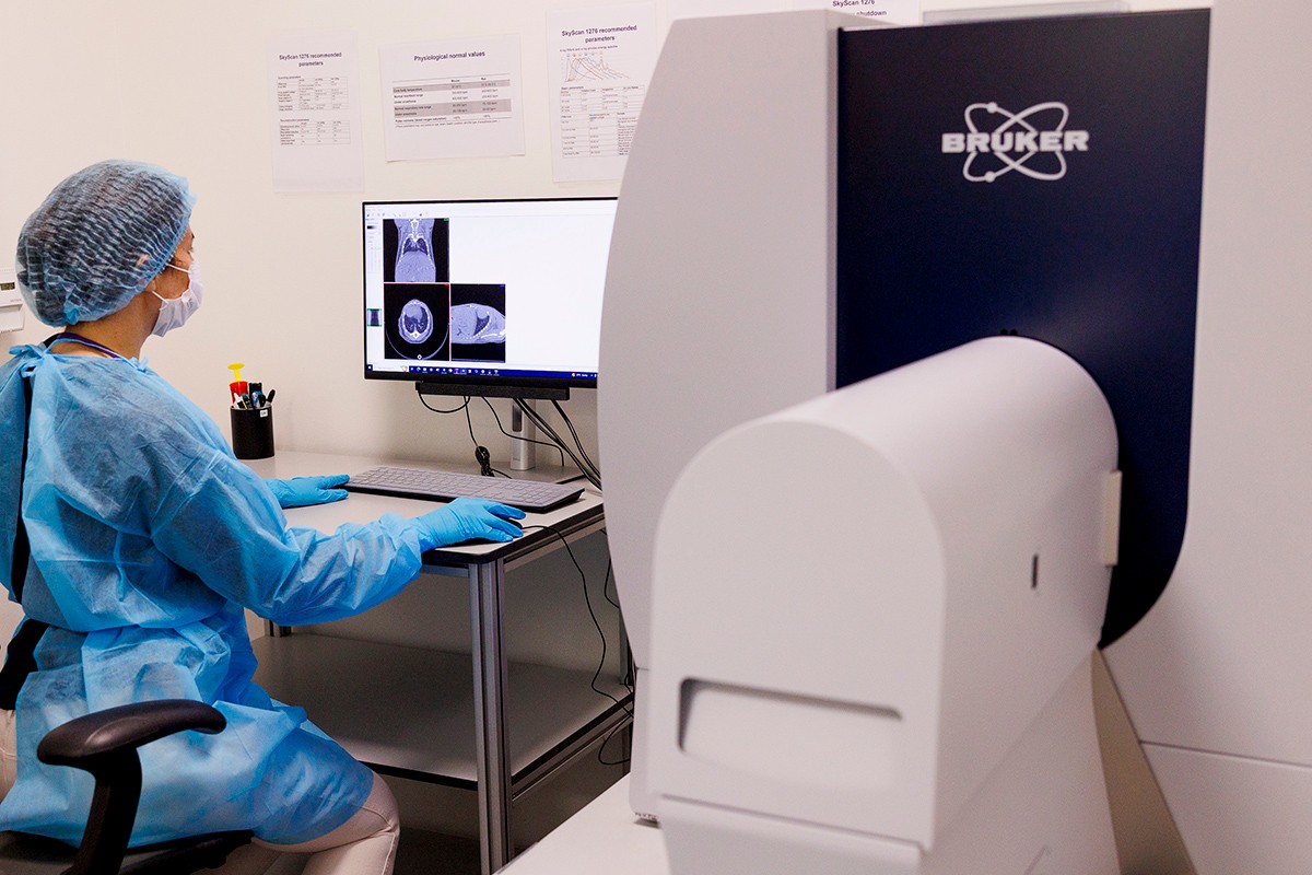
- Bruker SkyScan 1276 scanner for low-dose high-resolution micro-CT imaging enabling in vivo and ex vivo detailed structural and functional imaging
- Inscopix nVue-nVision platform for precise temporal correlations between behavior and neuronal activity
- Karl Storz Endo-Arthroflator-Vet endoscope for direct visualization of internal organs and biopsy procedures
- Ultima 2Pplus - Bruker- is a high-performance two-photon microscope for deep-tissue, high-resolution imaging of cellular and subcellular processes in living rodents
Supporting Equipment
- Anesthesia Machine for controlled live animal imaging procedures.
4. Computing Resources
The Biomedical Imaging Core is supported by a robust, high-performance computing infrastructure that includes a Dell PowerEdge server with 4 A100 GPUs, a dedicated XNAT database connected to the supercomputing cluster at NYUAD (Jubail), and specialized GPU-enabled processing workstations. This integrated environment leverages advanced analysis tools — such as BESA (for MEG and EEG), and AVISO for small animal visualization — to enable rapid deployment of deep learning applications across a variety of projects.