3D printers can make just about anything these days, from a pair of running shoes to chocolate, wood, and aircraft parts. And it turns out — even complex scientific gadgets used in cancer research.
Innovative, cost-efficient tools are essential in life sciences research to understand how cancer cells migrate from one place to another during metastasis, to probe how neurons connect in networks during human development, and to watch how white blood cells respond to infections. At NYU Abu Dhabi, biomedical engineers are designing new technologies biologists need to make important discoveries in these areas.
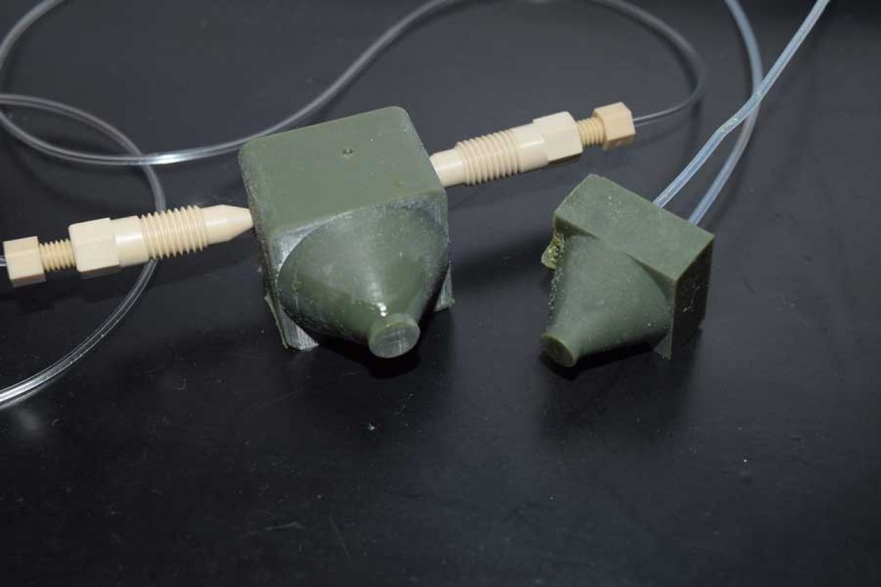
One such device is called a microfluidic probe, or MFP, as it's called in the lab. Typically made of glass or silicon, these very tiny scientific tools — roughly the size of a pen tip — were invented about a decade ago and are continuously being developed and refined. MFPs are used by scientists around the world to study, process, and manipulate live cell cultures in a controlled environment.
Although the technology is well established, it poses unique challenges and limitations. Specifically, MFPs cannot be easily produced on demand due to their complex fabrication procedures, and are expensive to make in large quantities because of their assembly procedures.
Enter the fascinating world of 3D printing.
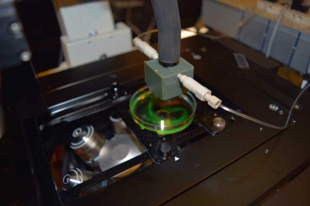
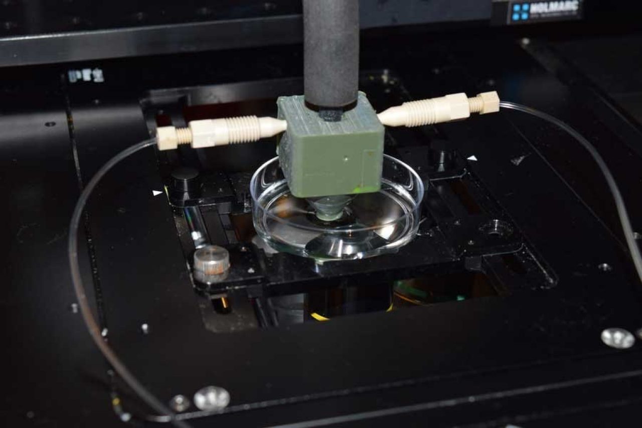
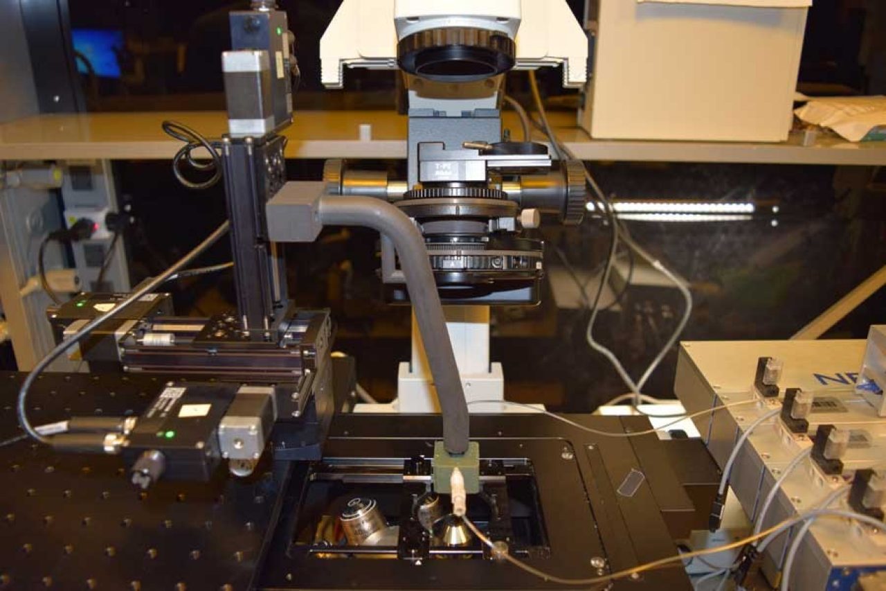
Photo Gallery: Images show a microfluidic probe printed in 3D. Cube-shaped and green-colored, this probe is about the size of a ring box, however, the design can be printed in many sizes.
“Democratizing” science technology
Biomedical engineers at NYUAD used a 3D printer to create a functional, integrated, and inexpensive MFP to study cancer cells and other living organisms in a Petri dish. Their printed device is cube-shaped with a cylinder tip and works with the same efficiency as its more expensive and laborious cousin.
“3D printers provide a simple, rapid, and low-cost technique for fabricating MFPs,” said Assistant Professor of Mechanical and Biomedical Engineering Mohammad Qasaimeh, whose team developed a framework to print microfluidic probes and quadrupoles in 3D.
“It’s cheaper to produce, easy to scale up or down, and fast to fabricate — all steps, from design to product, can be made in less than a day,” he explained, and as a result, “any science lab with a moderate resolution stereolithography printer will be able to fabricate 3D MFPs on demand and use them to process cells reliably.”
3D printed MFPs, “can deliver reagents in a localized manner, only a few tens of cells can be targeted within the culture dish, while leaving other millions of cultured cells untouched,” added Ayoola T. Brimmo, NYUAD Global PhD Fellow in Engineering and first author of the research, demonstrating its functionality in localized delivery and cell processing.


The study results, published in the journal Scientific Reports - Nature, builds on Qasaimeh’s previous work developing microfluidic probes and quadrupoles to study how human neutrophils (a type of white blood cell) behave when responding to infections.
In an earlier study, Qasaimeh and his research team used a silicon microfluidic probe to discover how neutrophils respond to moving sources of concentration gradients that mimic infections and pathogens. The research analyzed how quickly these cells respond to stimulation, showed how neutrophils start their migrations at a maximal speed that slows over time, and how neutrophils undergo rolling-like behaviours before they start to pursue an infection site.
Qasaimeh is the principal investigator of the Advanced Microfluidics and Microdevices Laboratory at NYUAD, whose work focuses primarily on developing micro-tools for biologists working in human health research, including devices to capture circulating tumor cells taken from blood samples of cancer patients.